Category: Lesions and moles


« Back to Blog

Caring for Your Skin, After Sun & Surgery

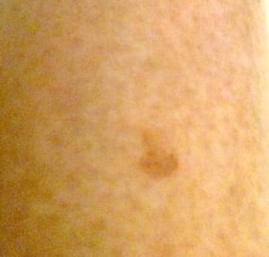
Taking care of your skin is crucial for effective healing after sun exposure and after surgery success. Summer months mean that skin damage from sun exposure is at its height. As summer comes to a close, it’s time to check your skin because detecting melanoma as early as possible makes it treatable. It is the deadliest cancer, but it doesn’t have to be if it it’s caught early. Regular self checking is a key part of immediate diagnosis is to do a regular self check.

AFTER SUN

What are you looking for? You’re at higher risk for melanoma if you’ve been sunburned frequently, have fair skin or a large amount of moles, live in a tropical or have a family history of melanoma. When you’re self checking, there’s a common acronym to help us remember what to look for. This is called the ABCDEs of warning signs for melanoma. This stands for:

  • Asymmetry: draw an imaginary line through the middle and it should look the same on both sides
  • Border irregularity: ragged or blurred, undefined edges
  • Color changes: a variety of colors within the same spot or mole
  • Diameter: greater than .25 inch (6 millimeters)
  • Evolving: changes over time

Where should you look? Important to look from head to toe, back and front. Using the angles from a hand mirror and a wall mirror to see the harder parts of the body from behind, literally start with the top of your scalp, especially if you haven’t worn hats to protect your head or have fine, thinning hair. Then move downward, examining the face, neck, shoulders, underarms, chest, torso, arms, hands, and nail beds. When checking the lower body, its easier to sit down, but don’t forget to check your buttocks, especially if you wear brazilian or thong bikinis. Look over your legs and feet, even the heels, soles of the feet and toenail beds.


AFTER SURGERY

What extra steps should I take? Whether your cosmetic procedure is invasive or non invasive, you should take some special care of your skin to promote healing and longevity of the effects of the procedure. Here are some simple tips to care for your skin after a cosmetic treatment:

  • Follow physicians directions. Each procedure has a unique protocol for optimal healing. This may include salves and the herbal supplement or cream version of Arnica Montana to reduce bruising.
  • Stay hydrated. Drinking plenty of water is good practice in general, but will help flush the area of toxins and empower healing. Avoiding alcohol and other dehydrating beverages so you don’t work against your natural healing processes.
  • Protect yourself from the sun. The skin is more vulnerable after a procedure. UV rays darken scars and this is what you want to prevent, so stay out of the sun.
  • Leave the recovery site alone. This means no picking or poking of the area. Disturbing the area could cause infection or unwanted scarring.
  • Use gentle skin care products. Make the extra effort during this time to pamper your skin while it’s healing. Use only mild, no fragrance skincare.

Help me to help you have beautiful, youthful skin by taking these precautions. From beginning to end, I look forward to helping you in the rejuvenation process. To schedule your consultation, visit www.davinciplastic.com.

Steven Davison M.D.

Board Certified Plastic/Reconstructive Surgeon

SOLUTIONS FOR KELOIDS…the SCARS THAT DON’T STOP!

SCARS  often come with a myriad of emotions.  Some scars remind us of pain, others of joy; of accidents or medical emergencies, or the miracle of ew life emerging via c-section deliveries.

In the eyes of a plastic surgeon, a scar is not our friend.  We want to leave the most minimal, if not invisible trace of our work, so a scar is not desirable.  In the case of keloids, I have the pleasure of helping to remove a scar, but not just any scar.  Keloids are scars that don’t stop!  They are not pleasant at all for the afflicted, and so it is personally rewarding to help individuals find relief from keloid scars.

WHAT ARE KELOIDS?   Keloids are scars that are usually discolored and raised.  They can be pink, flesh colored, or darkly colored in hues of red, purple and brown.  Keloids don’t blend in with the rest of skin texture since they’re often shiny, thick and fibrous.  They feel firm and rubbery to the touch, but can be uncomfortable.  They often cause severe itching and due to their thickness, can impair skin movement.  Keloids are not cancerous but result from an overgrowth of collagen type tissue at the site of a healed skin injury.  Here are some characteristics of Keloids:

  • extend past the borders of the original incision or cut into surrounding areas that weren’t injured                                                           shutterstock_72411928-1
  • irregularly shaped and tend to enlarge progressively
  • hard to treat and eradicate
  • can reoccur…even after year of being dormant
  • do not subside over time
  • typically appear following surgery or injury but can also appear due to slight inflammation, minor injuries or even spontaneously
  • can be triggered by burns, body or ear piercings, pimples, insect bites and simply scratching ones self

KELOID vs HYPERTROPIC SCARS … Skin’s normal healing process usually leaves a flat scar that fades over time.  Sometimes as the skin heals there appears a thicker scar, which could possible be a hypertropic scar.  Hypertropic scars exist only where the original wound was, although they may be more reddish and slightly raised.  They will blend in more with regular skin over time, although aggressive massage and steroid injections help them along.  To minimize Keloids, on the other hand, usually requires more treatment.

Here are some of the most “tried and true” treatments for Keloid removal:

  • Corticosteroid injected directly into the tissue once a month for about 3-4 months
  • Laser Excision followed by monthly intralesional steroid injections alone or in conjunction with Imiquimod
  • Surgical removal via scalpel, electric knife or laser for larger, more extensive formations

TREATING KELOIDS REQUIRE SKILL and EXPERIENCE…I have thousands of hours of surgical experience and specialized training.  Over the years, I have successfully been able to help many people who’ve suffered with Keloids.  I also take special care to offer complimentary post surgical treatments to minimize Keloid recurrence.

If you, or someone you  know is suffering with the discomfort of unsightly Keloids, let’s talk about the best options I can offer for your relief.

Dr. Steven Davison

Board Certified Plastic and Reconstructive Surgeon

Lipomas Removal…Quick Squeeze Method

Lipomas are the most common form of soft tissue tumors and they are formed with adipose (fatty) tissue. Lipomas can form in many shapes, sizes and places on the body and they occur more often during middle age (40 to 60) but they are often diagnosed in children as congenital lipomas. They are soft to the touch, moveable and usually painless. Typically they grow just between the skin and underlying muscle but can also be found in the deeper muscle tissues such as the intermuscular septa, or in organs like the abdominal organs, the oral cavity, the internal auditory canal, the cerebellopontine angle and the thorax. They require treatment by a plastic surgeon for safe, effective removal. Lipomatosis, also called liposis, adiposis or lipomatoza and simply refers to groups of lipomas in one location on the body.

Other names for lipomas (types) include:

  1. Adenolipomas are associated with the sweat glands
  2. Conventional lipomas are the most common type composed of mature white fat
  3. Hibemomas are made up from brown fat
  4. Fibrolipomas are fat and fibrous tissue
  5. Angiolipomas are mostly blood vessels and fat tissues
  6. Myelolipomas are fat and the tissues that make blood vessels
  7. Spindle Cell lipomas are rod shaped, seen mostly in elderly men
  8. Intradermal spindle cell lipomas is most common in women
  9. Pleomorphic lipomas are the various shapes and sizes
  10. Atypical lipomas are composed of a large number of “deeper” fat cells
  11. Neural lipomas are fibrous, fatty tissues
  12. Others include: Proteus Syndrome, Cowden Syndrome caused by PTEN gene mutations, Familial lipodystrophy, Hibernomas and Familial angiolipomatosis.

METHODS OF REMOVAL

Lipoma cases are nearly always referred to a surgeon who is skilled and qualified in their removal.

Typical Treatments:1) endoscope assisted removal: 2) suction assisted removal, 3) minimal incision removal and 4) remote incision lipoma extraction.  These all work and have been used by surgeons for decades to remove lipomas of all types.  But, some of these techniques are tedious and require extra equipment (even though they are advanced treatments) and are not always necessary since a quicker, simpler method is now in use.

I helped pioneer a very simple, safe and effective method that involves a “simple incision and a quick squeeze” to remove lipomas without all the extra instruments used in other methods.

Once surgeons learn the quick squeeze method and start using it, they realize it’s great benefits. It’s safe, quick, simple and effective.

QUICK SQUEEZE LIPOMA REMOVAL

1) The lipoma is identified by palpation and its margins are outlined.  2) The area is then infiltrated with local anesthesia and is prepared and draped in standard fashion.  3) An incision approximately one quarter to one third the diameter of the lipoma is made (carried down to the lipoma capsule).  4) The lipoma is then squeezed out through the incision…without any additional dissection.


LIPOMA STUDY

Involved 140 lipomas removed, in 50 patients, over the life of this study.These lipomas were located on the face, scalp, neck, thorax, back and flanks, hips/pelvis and on both the upper and lower body extremities.  Of the 140 removals, there were only two recurrences, no complications, no revisions.

Simple Incision, Quick Squeeze technique for lipoma removal makes them gone in 30 seconds!

See complete information about this method of lipoma removal at: www.DAVinciplasticsurgery.com.

Dr. Davison


KELOIDS…require expert attention to deal with the pain, itching and physical disfigurement.

KELOIDS are a type of scar that results from an overgrowth of collagen type tissue at the site of a healed skin injury.  Keloids can be firm, rubbery lesions or shiny, fibrous nodules that vary in color….some are pink, others flesh colored or they may be red to dark brown in color.  Keloids are benign (not cancerous), are not contagious but often cause severe itching and changes in skin texture.  Severe keloids can affect movement of the skin.

Keloids are often confused with “hypertropic scars.”  They are not the same. Hypertropic scars are raised and they do NOT grow beyond the boundaries of the original wound.

Since I have specialized training and years of experience in surgery and reconstructive surgeries, I have dealt with so many patients in need of Keloid removal and scar revision…I couldn’t begin to number them.  Just know, that dealing with Keloids is serious business and you should see a surgeon with expertise in this area…in order to get desirable results.

KELOIDS…WHERE THEY OCCUR ON THE BODY...Keloids can develop anyplace where an abrasion (injury) has occurred.  They can even result from pimples, insect bites, scratching ones self, from burns or other skin trauma.  They can develop post surgery.  They most commonly occur:

  • On the central chest
  • The back & shoulders
  • On Ear lobes
  • On arms & over the collar bone after body piercings
  • From shaving…razor bumps may become keloids
  • Bottom of feet
  • Can occur at any age…but generally don’t until past age 11
  • Keloids…may be linked to hereditary tendencies

COMMON TREATMENTS FOR KELOID REMOVAL

1)  Corticosteroid injected directly into the tissue – typically 3-4 treatments spaced about 1 month apart – will reduce a raised Keloid to a flat scar and reduce redness, itching and burning.

2)  Surgical Removal is used for larger, more extensive formations.  Technique involved may employ a scalpel, electric knife or laser removal.

3) Post-Surgical Complimentary Treatments are often used to minimize Keloid recurrence after surgical removal.  These treatments include:   -silicone sheet dressings applied directly to the skin around the incision; -compression therapy the mechanically flatten the scar formation, – topical imiquimod or advanced injectables such as bleomycin,  -interferon or 5-fluorouracil.  These drugs work at the cellular level to disable the Keloid tissue formation process.

4)  Laser Excision followed by monthly intralesional steroid injections alone or in conjunction with imiquimod.

These are my treatment preferences…even though others may exist.  I stay with the tried and true…those options proven to get desirable results.

OK….last two blogs have covered skin problems….lipomas and keloids.

Next blog I’ll move back into FEMGen options….today they are no longer “taboo.”  Women are lining up to have life changing treatments that restore their confidence, youthfulness, that rev up their love lives…and restore health and well being in many cases.  Don’t suffer any longer in silence.   Women everywhere are stepping up and speaking out….the embarrassment is gone, Designer Vaginas are in and complete health and vitality for the “V Zone” is here to stay! 

More on that next blog!

Stay Well.

Steven Davison M.D.

DAVinci….where I combine the supreme elements of art and surgical science.”

Lipomatosis…nobody wants it….many people get it…one simple “squeeze technique” can remove it in 30 seconds!

LIPOMATOSIS…is the presence of multiple lipomas on the body.  Lipomas are adipose tumors….these are tumor growths filled with mature fat cells.  Lipomatosis is also referred to as: liposis or adiposis.  Whatever it is called…it simple means an abnormal localized tumor like accumulation of fat in the tissues.  Many discrete, encapsulated lipomas form on the body trunk and extremities with very few on the head.  In 1993, a genetic defect within lipomas was localized to chromosome 12q15, where the HMGIC gene encodes the high-mobility-group protein isoform 1-C.  This is one of the most commonly found mutations in solitary lipomatous tumors, while lipomas often have multiple mutations.  Other chromosomes have also been observed.

Other entities often accompanied by multiple lipomas include Proteus syndrome, Cowden syndrome and related disorders due to PTEN gene mutations, benign symmetric lipomatosis (Madelung disease), familial lipodystrophy, hibernomas and familial angiolipomatosis.  Below I outline some symptoms….because more people are presenting today with lipomas….it is important to educate on this subject.

SYMPTOMS of FAMILIAL LIPOMATOSIS (FML)…FML is a rare disorder that tends to run in families and affects both men and women.  It is hereditary and shows up as groups of fat cells in the subcutaneous tissues that begin to grow and produce multiple fatty lumps of “benign” fatty tumors (lipomas).

  • Usually appear on the abdomen, arms, thighs, neck
  • Can also appear on the face, buttocks, lower legs
  • Tend to vary widely in size…& may or may not be surrounded by connective tissue
  • May or may not be attached to the skin & nearby muscle
  • They are rubbery, moveable & ovoid to round in shape
  • Are not usually painful (unless in an irritated area)
  • Can appear singularly or in clusters
  • Typically appear during of soon after adolescence
  • Extreme cases can grow to appear grotesque & can interfere with mobility, movement & cause disfigurement

LIPOMA REMOVAL REQUIRES THE SKILLS OF A QUALIFIED SURGEON…LIPOMA cases are nearly always referred to a surgeon who is skilled and qualified in their removal.

My previous blog covered methods of Lipoma’s removal…surgical excision, endoscopic removal, liposuction and a simple “squeeze” technique I have developed that safely and effectively removes many lipomas within just seconds through a small incision.

LIPOMATOSIS….NOBODY WANTS IT…WE CAN DEAL WITH IT!  If you, a friend or loved one has been suffering with Lipomas.  Please know there is help!  Come in and let’s get your treatment plan going so you can be free again.

OK….MY NEXT BLOG WILL COVER KELOIDS….THEN WE’LL GET OFF THIS TRACK AND GET BACK INTO BEACH BODY TOPICS!

HOPE EVERYONE IS HAVING A WONDERFUL SPRING!

Steven Davison M.D.

“DAVinci….where I combine the supreme elements of art and surgical science.”

IS IT THIS ONE…OR IS IT THAT ONE?

A quite frequent problem involved in the removal of small lesions such as moles, nevi and other lesions is…correct identification.  Correct identification and confirmation of the exact site for these small lesions is paramount.  But often, this is a problem when a patient is referred from a dermatologist and presents in our office.

Identification and Clinical Notes The most common way of locating and identifying the correct site is by asking the patient to identify the site in conjunction with clinic notes.  This method is used to help maximize accuracy in the ID process.  Problem is…this can backfire.

I’ve actually had a patient blow up at me when I asked them to confirm which mole the dermatologist wanted biopsied.  But there is an easy solution to this problem.

Smart Phone’s High Pixel Camera! The Smart Phone’s high pixel camera does not require additional lighting, so you can take a photo in the dermatologist’s office without hassle.  Here are the steps I recommend:

  1. Take a photo (or have someone on staff do it)…of the marked lesion.
  2. Include a frame of reference – e.g. the nose, right ear, left ear, belly button – something discernible.
  3. If the lesion is hard to find, include a ruler located by the lesion so we can measure from the landmark.
  4. This prevents hours of anguish caused from having to revisit the dermatologist and from incorrect lesion biopsy mistakes.

Be thankful for modern technology…quite amazing!

Happy Thanksgiving!

Dr. Steven Davison

Schedule Your Consultation With Dr. Davison

The initial consultation with your surgeon is critical! It should always be face to face with your surgeon during which time he/she listens closely to your concerns. Dr. Davison believes this time spent with you is critical to both you and to him. It is during the consultation that he comes to fully understand your concerns, evaluates them correctly and is then able to make the recommendations and develop the treatment plan thats right for you. Dr. Davison invests the time to sit down and talk to you face-to-face.

  • This field is for validation purposes and should be left unchanged.

3301 New Mexico Ave. Suite 236

Washington D.C.

Schedule An Appointment